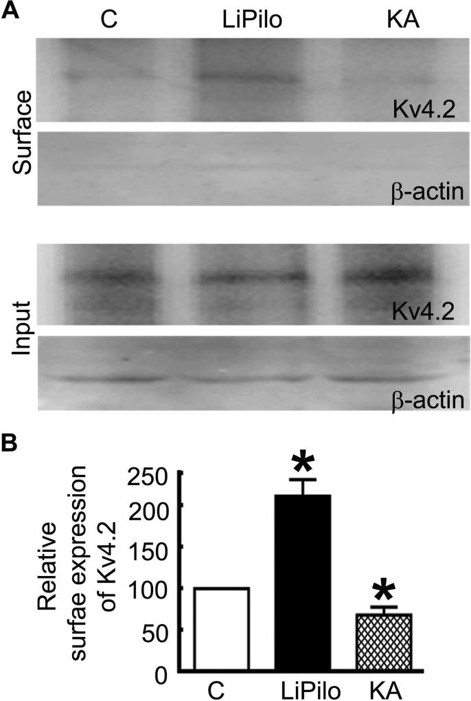
Figure 4. The surface expression of Kv4.2 was increased during LiPilo-induced SE but reduced during KA-induced SE.
A, Sample Western blots of the surface protein fraction and the total protein fraction (input) of the potassium channel Kv4.2 in hippocampal slices acutely obtained from P23-25 control animals (C), animals in status epilepticus of 1 hour in duration induced by a combination of lithium and pilocarpine (LiPilo), and animals in status epilepticus of 3 hours in duration induced by kainic acid (KA). The purity of surface proteins was checked in each assay by confirming the absence of β-actin expression in the surface protein fraction. B, The surface expression of the Kv4.2 subunit presented as mean ± SEM of the ratio of the surface-to-total expression in slices obtained from control animals (C), LiPilo-SE-treated animal (LiPilo), and KA-SE-treated animals (KA). n=6 each, * p < 0.05.

