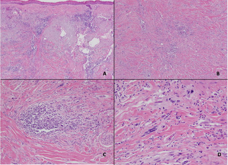
Figure 1.

Desmoplastic melanoma. Microscopic images of a desmoplastic melanoma excision showing a low-power view of an effaced epidermis overlying sun-damaged skin containing an invasive, infiltrative spindle cell proliferation with scattered intratumoral lymphoid aggregates (A, H&E 2x). Deep within the dermis, the spindled melanocytes are seen within a desmoplastic stroma (B, H&E 4x) with scattered lymphoid aggregates among the malignant cells (C, H&E 10x). Cytologically, the melanocytes display significant pleomorphism and mitotic activity (D, H&E 20x).
