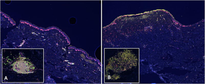
Figure 4.

Tertiary lymphoid structures (TLS) identified in scars by multispectral immunofluorescence imaging (spectrally unmixed, 20x). Low-power images show the brightly staining lymphoid aggregates in the papillary dermis. Inset images highlight a single TLS. (A) A hypertrophic scar collected from an excision of an atypical melanocytic lesion. (B) A scar excised from the site of a junctional nevus with melanocytic hyperplasia.
