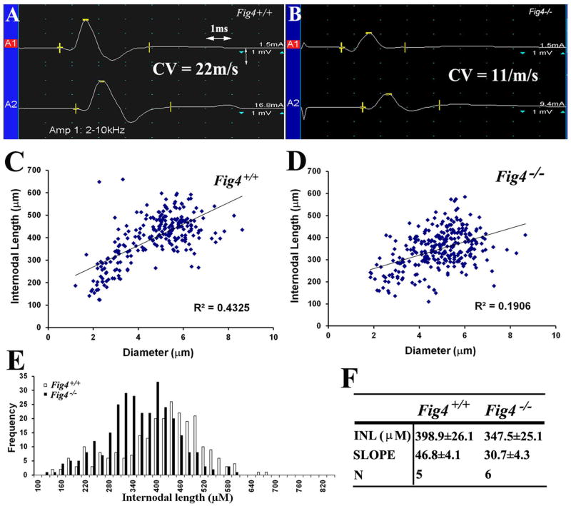
Figure 3.
(A). Compound muscle action potential (CMAP) was recorded from a p21 Fig4+/+ mouse paw. The motor nerve conduction velocity (CV) was 22m/s. (B). The same experiment was done in a p21 Fig4−/− mouse and showed a 50% reduction of CV. (C–D). Mouse sciatic nerves were teased into individual fibers in liquid Epon and imaged. Internodal length and diameter were measured and plotted. The slope in Fig4−/− mice was lower than that in Fig4+/+ mice, supporting shortened internodal length in Fig4−/− mice. (E). The frequency vs. diameter plot allows a better visualization of shortened internodes mainly involving those longest (largest) fibers. (F). INL = internodal length; N = number of mice; p<0.05 for the comparison of both internodal length and slope.

