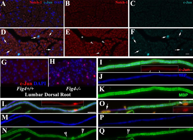
Figure 4.
(A–F). A, B and C are for Fig4+/+ nerves. D, E and F are for Fig4−/− nerves. Transverse sections of adult mouse sciatic nerves were stained with antibodies against c-Jun and Notch-1. In line with previous studies, Notch-1 was localized in myelin (A, B, D, and E). F. There was an increase of c-Jun in the Schwann cell nuclei of Fig4−/− nerves, compared with that in Fig4+/+ nerves in C. (G–H). There were numerous nuclei in Fig4−/− spinal roots showing strong c-Jun signal, which was hardly seen in Fig4+/+ spinal roots. (I–K). A wild-type myelinated nerve fiber from a sciatic nerve was stained with antibodies against c-Jun and MBP (which labels myelin). The axon was labeled with YFP (artificially changed to blue color) expressed under a neuronal specific promoter. c-Jun staining was hardly visible in the nucleus (arrowheads in inset). (L–N). A Fig4−/− sciatic nerve fiber with subtle demyelination (between arrowheads in N) showed a robust increase of c-Jun (inset in L) in the Schwann cell nucleus. (O–Q). The increase of c-Jun (inset in O) was seen in a Fig4−/− sciatic nerve fiber with severe segmental demyelination (left to arrow in Q). The Schwann cell nucleus at the middle of the internode (arrowhead in O and inset in O) showed an increased c-Jun. Another nucleus in the demyelinated region also expressed a high level of c-Jun (arrow in O). Scale bars = 10µm

