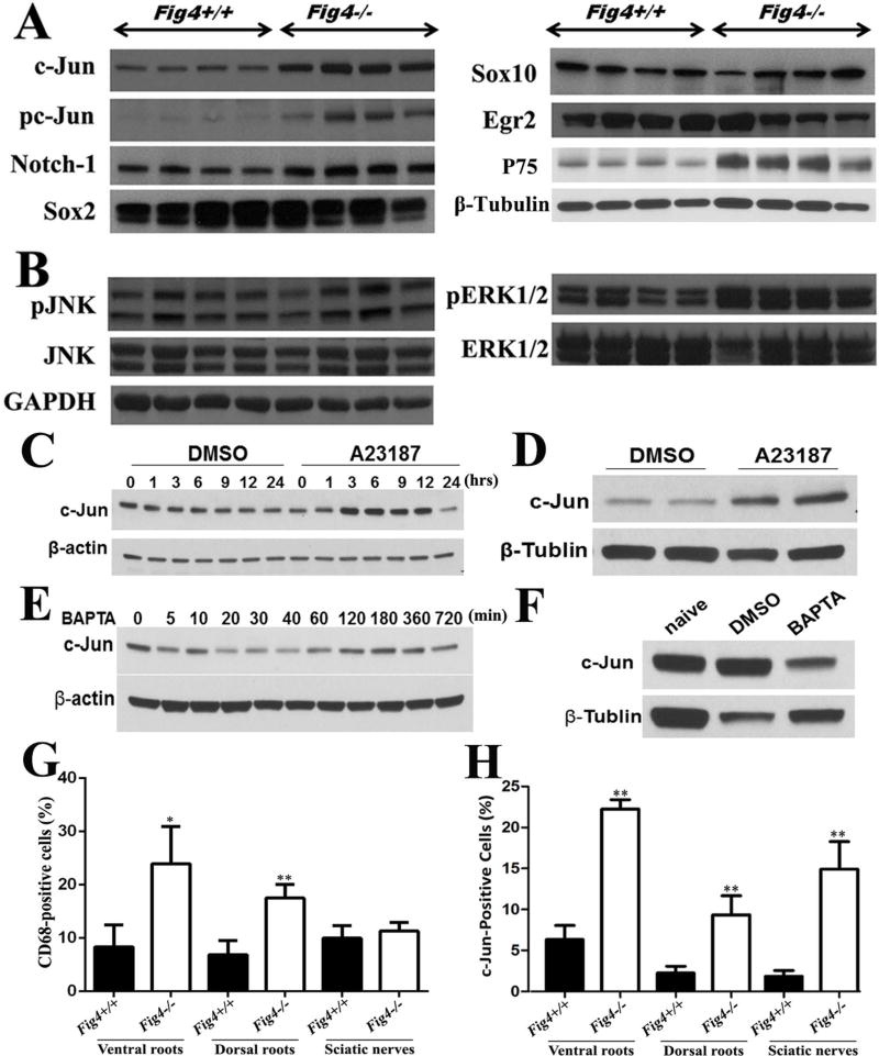
Figure 5.
(A). Mouse sciatic nerves at the age of p21 days were studied by Western blot. c-Jun, p75 and phosphorylated c-Jun (pc-Jun) were robustly increased in Fig4−/− mice. Notch-1 was marginally increased (p=0.048). Sox10 and Egr2 did not show significant changes in Fig4−/− mice. (B). Molecules up-stream and down-stream to c-Jun were also evaluated in Fig4−/− mice. GAPDH = loading control. (C). Schwann cells in culture were treated with Ca2+ ionophore A23187 (2.5µM). c-Jun levels were increased after 1 hour. There were numerous cells dead at 24 hours, leading a decrease of c-Jun level. (D). Sciatic nerves in wild-type mice were surgically exposed and wrapped by gauzes soaked with 19µM A23187 for 4 hours. Nerves were then dissected for Western blot analysis to measure c-Jun levels. (E). Schwann cells were incubated with BAPTA (30µM). Protein lysates were extracted from the cells at different time points for Western blot. (F). Two wild-type mice were treated with either vehicle or BAPTA (5mg/kg; i.p. daily) for 7 days. A naïve mouse was used for control. Sciatic nerves were then collected for Western blot analysis. (G). Transverse section of paraffin embedded mouse nerves at p21 of age was stained with antibodies against CD68. The increase of CD68 was robust in spinal roots of Fig4−/− mice but was not seen in the sciatic nerves. (H). When c-Jun was stained, its increase was found in all parts of peripheral nerves. * = P< 0.05, ** = P< 0.01

