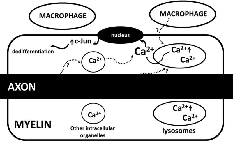
Figure 7.
A proposed mechanistic model to be further tested in the future: Release of Ca2+ from lysosome is primarily through two Ca2+ channels, TRPML1 and TPC245. PI3,5P2 binds with TRPML1 to activate Ca2+ efflux through TRPML110. Since PI3,5P2 is deficient in Fig4−/− cells, lysosomes develop a high level of Ca2+. The extra Ca2+ in Fig4−/− lysosomes can be released by internal or external signals (speculated from axons or macrophages) to stimulate expression of dedifferentiation molecules like c-Jun, promoting demyelination. Excessive Ca2+ could also promote demyelination via other unidentified pathways.

