. 2018 Apr 16;12(2):263–271. doi: 10.4184/asj.2018.12.2.263

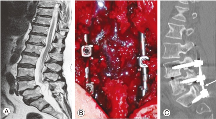
Fig. 2. Pre-, intra-, and postoperative imaging of patient 21 of Table 1. This patient was a 74-year-old female with 6 months??history of L5 bilateral radiculopathy and claudication. (A) Preoperative sagittal T2-weighted magnetic resonance imaging showing L4–S1 stenosis with the L4–L5 pathological disc (Pfirmann 4) at the same level as that of the planned laminectomy with first-grade spondylolisthesis and a caudal pathological disc (Pfirmann 4). (B) Intraoperative picture of a three-level stabilization (L4–L5–S1) with the hybrid device (rigid L4–L5 and dynamic L5–S1). (C) Postoperative computed tomography with sagittal reconstruction demonstrating the correct positioning of the device and dynamic segment of stabilization (white arrow).

Fig. 2