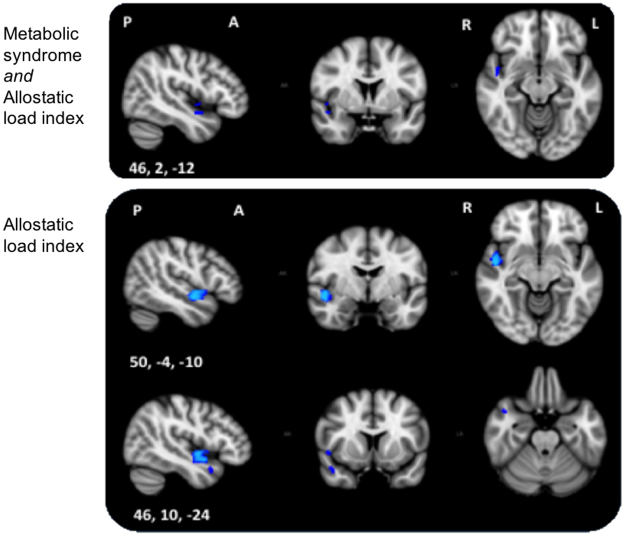
Figure 2.
Top: F-test 1 of unique metabolic syndrome or allostatic load index association with grey matter after removing the effects of Framingham stroke risk and socio-demographic variables. Significant results extend the right insular and opercular cortex. Bottom: Post-hoc t-test shows an association between allostatic load index and lower voxelwise grey matter after removing the effects of metabolic syndrome, Framingham stroke risk and socio-demographic variables. Significant voxels were located along the right insular and opercular cortex, superior temporal gyrus and temporal pole. Blue represents regions significant at p < 0.05, threshold-free cluster enhancement, multiple comparisons corrected. P, posterior; A, anterior; R, right; L, left; Coordinates are in MNI space.

