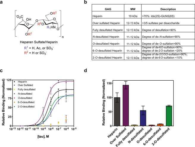
Figure 3.
Binding of tau to heparin derivatives. (a) HS chains consist of GlcA/IdoA-GlcNAc disaccharide units that can be modified at positions indicated in red and blue. (b) List of heparin/heparin derivatives that were used, their average molecular weights, and description of their sulfation modifications. (c) Binding of 2N4R to various heparin derivatives by ELISA with data fit to a Hill binding model (line) where appropriate. (d) Normalized relative binding of 2N4R (10 nM) to various heparin derivatives. Three independent experiments were performed, data were normalized to heparin controls, and the error shown is SD.

