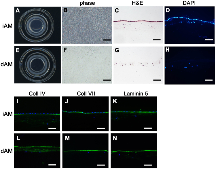
Figure 1.
Macroscopic and microscopic evaluation of iAM and dAM. Cryopreserved iAM mounted on the culture insert showed sub-transparent appearance (A). Phase contrast image of the iAM whole mount showed uniformly arranged and well maintained epithelial cells (B). H&E (C) and DAPI staining (D) of iAM crosssection showed organized epithelial layer. Transparent dAM after 0.02% EDTA treatment and electric toothbrush scraping (E). Phase contrast image of the dAM whole mount showed few epithelial cells (F). H&E staining (G) and DAPI staining (H) of dAM crosssection confirmed the presence of an intact stroma without the epithelial layer. Immunostaining of collagen IV (I,L), collagen VII (J,M) and Laminin 5 (K,N) showed all these basement membrane components were well preserved after amniotic membrane epithelial denudation. Bars represent 500 μm in (B and F), and represent 100 μm in (C,D,G,H, I–N).

