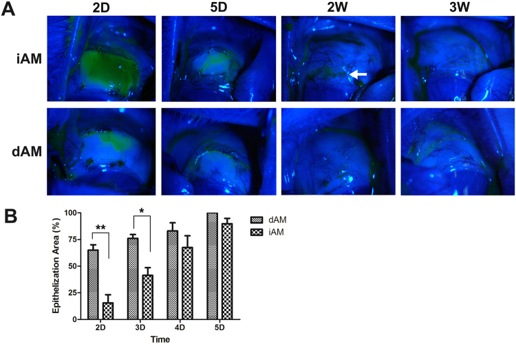
Figure 3.
Evaluation of epithelialization using fluorescein corneal staining. Two days postsurgery, the iAM surface showed large area of fluorescein staining, whereas on dAM surface the fluorescein uptake was much smaller (2D). Five days after surgery, the central area of iAM surface showed positive staining, while the dAM surface was almost negative (5D). Persistent dots fluorescein staining was observed on iAM even after 2 weeks (2W, arrow), while was completly negative on the dAM (A). Statistical analysis showed the area of epithelialization on dAM vs iAM, there was significant difference in day 2 and day 3 (B, *p < 0.05, **p < 0.01).

