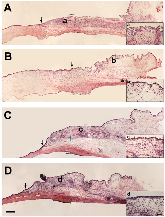
Figure 4.

H&E staining on rabbit corneal and conjunctival tissue after AM transplantation. Visible stratified epithelium was observed 2 weeks post surgery with iAM (A) and dAM (B). Uniform epithelial layer and intensified stromal cellularization was observed in iAM group 3 weeks post surgery. In addition, the stroma showed folded dense matrix like embedded structure (C, blue arrow head). Three weeks post surgery sections of dAM transplanted group showed smooth and homogenous stroma (D). Inserted figures show high magnification of the dotted portion (a,b,c,d). The black arrow heads show the corneal limbal junction. Bar represents 500 μm in (A–D), and represents 100 μm in a,b,c, and d.
