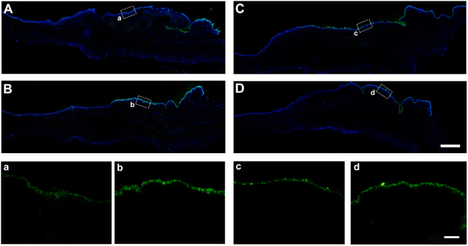
Figure 7.
K19 immunostaining on ocular surface epithelium after AM transplantation. Two weeks after iAM (A) or dAM (B) transplantation, K19 positive staining was specific to the conjunctival surface of the transplantation area in both groups, the epithelium of the limbal area was K19 negative. High magnification images showed K19 expression was stronger on dAM group (b) than that on iAM group (a). Three weeks after iAM (C,c) or dAM (D,d) transplantation, K19 expression showed similar pattern without any significant difference. Bar represents 500 μm in (A–D), and represents 100 μm in a–d.

