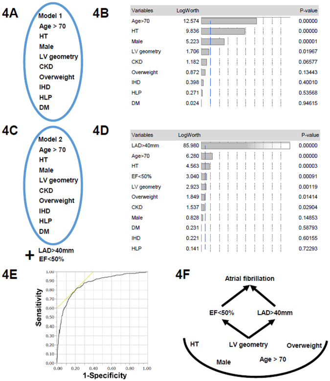
Figure 4.
(A) Model 1 in Table 3 includes factors included age >70 years; sex; LV geometric remodelings that were defined as ordered variables (normal geometry, concentric remodeling, concentric hypertrophy, and eccentric hypertrophy); and the presence of comorbidities such as ischemic heart disease (IHD), hypertension (HT), diabetes mellitus (DM), hyperlipidemia (HLP), CKD and overweight (body mass index >25 kg/m2). (B) LogWorth scales of multivariate model 1 in Table 3. The p-values were transformed to the LogWorth (−log10(p-value)) scale. Hence, any LogWorth above 2 corresponds to a p-value below 0.01. A LogWorth of zero corresponds to a nonsignificant p-value of 1. (C) Model 2 includes factors included as factors for model 1, as well as left atrial enlargement [LAD (left atrial diameter) >40 mm] and low LVEF (EF <50%). (D) LogWorth scales of multivariate model 2. (E) Results of receiver operating characteristic (ROC) curve analysis. For predicting the presence of AF, the formula was generated from a linear regression (x = 3.55296 + 0.0208 × LVEF +− 0.09732 × LAVI). The prediction formulas for the response levels predicting the presence of AF and the absence of AF were (1/(1 + Exp(x)) and (1/(1 + EXP(−x)), respectively. Then we compared the two levels and predicted the presence or absence of AF according to the larger response level. The ROC curve shows an area under the curve of 0.871, and when x = 2.1433, the sensitivity is 0.8044 and 1-specificity is 0.6042. (F) A proposed schema for the relationship between LV geometry, LVEF, LA enlargement, and AF.

