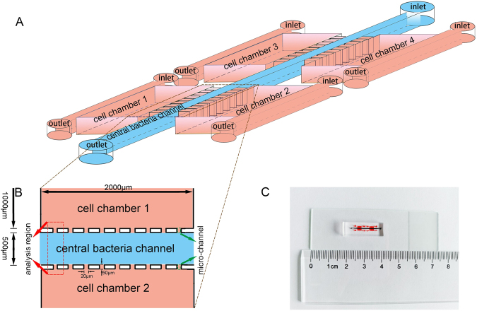
Figure 1.
Design and illustration of the microfluidic device. (A) A schematic diagram of the device for studying bacterial chemotaxis mechanism. Microfluidic device comprised of four separated cell culture chambers, one central bacteria channel and micro-channels between them. (B) The analysis region was sited between two chambers of cell culture, where the preferential accumulation of bacteria could be quantified. Micro-channels were the barriers between the central bacteria channel and the cell chambers, and also acted as flow regulators. (C) A photographic overview of the integrated microfluidic device.

