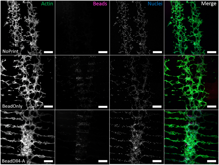
Figure 4.
Microscopic immunofluorescent images of labelled sprouting HUVECs, in Matrigel enriched media, on top of gelatin coated glass (NoPrint, top row). Sprouting HUVECS in Matrigel enriched media on top of a line pattern of fluorescent IgG beads coated with gelatin (BeadOnly, middle row) or on a Dll4 ligand immobilized with fluorescent IgG beads line pattern coated with gelatin (BeadDll4, bottom row). First three columns contain intensity values separately. The last column contains the merged image with in green; actin stained with phalloidin, in pink; fluorescent IgG beads, and in blue; nuclei stained with DAPI. Scale bar represents 250 µm.

