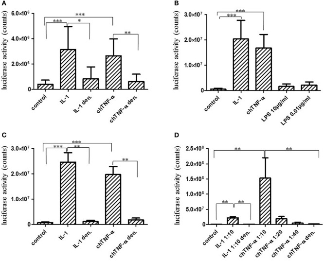
Figure 7.
Biological activity of chicken TNF-α (chTNF-α). The full-length TNF-α gene was expressed in HEK293 cells and concentrated cell culture supernatants were added to CEC–NFκB–LUC reporter cells at a 1:25 dilution for 6 h. Biological activity was quantified using a luciferase reporter assay. Luciferase activity is expressed as counts. COS cell expressed chicken interleukin (chIL)-1 was used as a positive control (38). Cytokine preparations were denatured (den.) by heating to 80°C for 5 min. Results represent three independent experiments (A). Full-length chTNF-α (B,C), and the extracellular domain of chTNF-α (D) were expressed in E. coli and purified by affinity chromatography. Biological activity was quantified as described in (A). E. coli lipopolysaccharide (LPS) was added to the reporter cells in parallel to test their sensitivity to potentially contaminating LPS (B). E. coli expressed chTNF-α or control chIL-1β were heated to 80°C for 5 min to inactivate the protein (C). Reporter cells were activated with different dilutions of the extracellular domain of chTNF-α and the biological activity quantified as above (A). Results shown in (A–D) each represent three independent experiments. *p ≤ 0.05; **p < 0.01; ***p < 0.001 (Mann–Whitney U-test, Student’s t-test).

