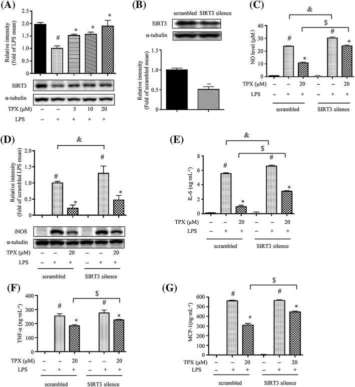
Figure 3.

The anti‐inflammatory effect of TPX in RAW264.7 macrophages is partially mediated through SIRT3. (A) The protein expression of SIRT3 was determined by Western blotting (n = 6). α‐Tubulin was used as an internal loading control. Data were normalized to the mean value of the LPS group. (B) SIRT3 expression in SIRT3 silenced RAW264.7 macrophages (SIRT3 silence) and control cells (scrambled) was determined by Western blotting (n = 6). α‐Tubulin was used as an internal loading control. Data were normalized to the mean value of scrambled group. (C) Cells were treated with 20 μM TPX in the presence of LPS (1 μg·mL−1) for 18 h. NO production was determined by Griess reagent (n = 6). (D) iNOS abundance was measured by Western blotting (n = 6). Data were normalized to the mean value of the scrambled LPS group. Cells were pretreated with TPX for 1 h and then stimulated with LPS (1 μg·mL−1) for 18 h. The levels of IL‐6 (E), TNF‐α (F) and MCP‐1 (G) were determined by ELISA kits (n = 6). Data are expressed as means ± SEM. # P < 0.05 versus DMSO, * P < 0.05 versus LPS, & P < 0.05 versus scrambled LPS, $ P < 0.05 versus scrambled TPX.
