
Figure 4.
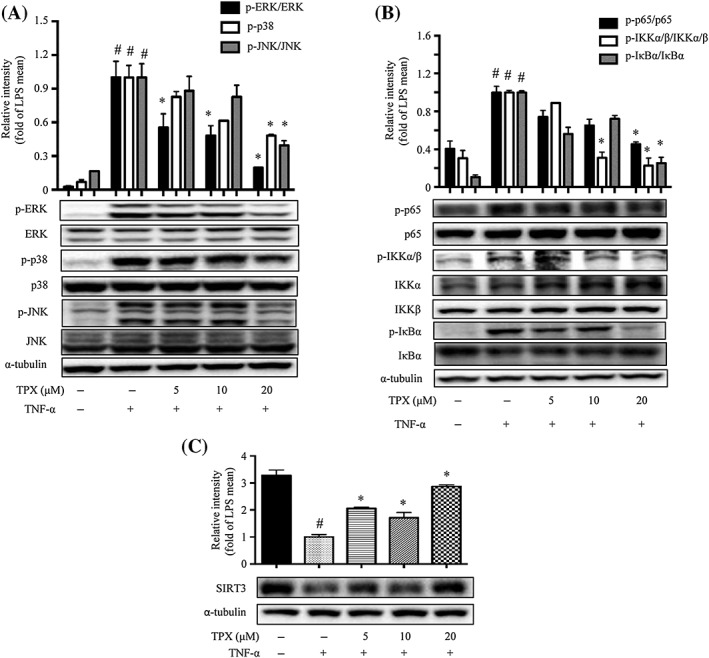
TPX prevented TNF‐α‐induced inflammatory changes in adipocytes. Differentiated 3T3‐L1 adipocytes were treated with TPX for 6 h and subsequently stimulated with TNF‐α (15 ng·mL−1). (A) The protein levels of phospho‐ERK, ERK, phospho‐p38, p38, phospho‐JNK and JNK were detected by Western blot analyses (n = 6). α‐Tubulin was used as an internal loading control. Data were normalized to the mean value of the TNF‐α group. (B) The protein levels of phospho‐IKKα/β, IKKα, IKKβ, phospho‐IκBα, IκBα, phospho‐p65 and p65 were detected by Western blot analyses (n = 6). α‐Tubulin was used as an internal loading control. Data were normalized to the mean value of the TNF‐α group. (C) The protein expression of SIRT3 was determined by Western blotting, and α‐tubulin was used as an internal loading control (n = 6). Data were normalized to the mean value of the TNF‐α group. Data are expressed as means ± SEM. # P < 0.05 versus DMSO, * P < 0.05 versus TNF‐α.