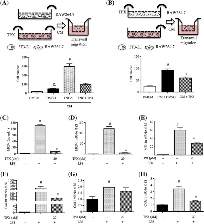
Figure 5.

TPX decreased the migration of RAW264.7 macrophages towards 3T3‐L1 adipocytes. (A, B) Procedures of the macrophage migration experiments. (A) Macrophage migration assays using DMEM (migration media) or CM from adipocytes treated with vehicle, TNF‐α (15 ng·mL−1) or TNF‐α (15 ng·mL−1) + TPX (20 μM) for 24 h. Migrated RAW264.7 macrophages were visualized by DAPI staining and quantified (n = 6). (B) RAW264.7 cells were treated with vehicle or TPX (20 μM) for 4 h, and the detached cells were used for the migration assay in the presence of DMEM or CM. Migrated RAW264.7 macrophages were visualized by DAPI staining and quantified (n = 6). (C) Cells were pretreated with TPX for 1 h and then stimulated with LPS (1 μg·mL−1) for 18 h. The levels of MCP‐1 were determined by an ELISA kit (n = 6). RAW264.7 macrophages were pretreated with TPX (20 μM) for 1 h and then stimulated with LPS (1 μg·mL−1) for 6 h. The mRNA levels of MCP‐1 (D), MIP‐1α (E), Cxcl10 (F), Ccl11 (G) and Cx3cl1 (H) were analysed by real‐time RT‐PCR, and normalized to 18S (n = 6). Data are expressed as means ± SEM, # P < 0.05 versus DMSO, * P < 0.05 versus LPS, & P < 0.05 versus DMEM.
