Figure 1.
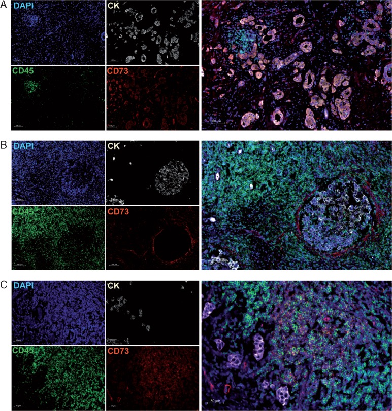
CD73 expression in TNBC. Representative images of the immunofluorescence staining with DAPI (blue online), cytokeratins (white online), CD73 (red online) and CD45 (green online) on TNBC tissues. Scans were imaged at 200× magnification using Zen lite software (Carl Zeiss). (A) CD73 is expressed on tumor cells and in the peri-tumoral stroma. (B) CD73 is expressed on few CD45+ leukocytes and stromal cells. (C) CD73 is expressed mainly on CD45+ leukocytes.

