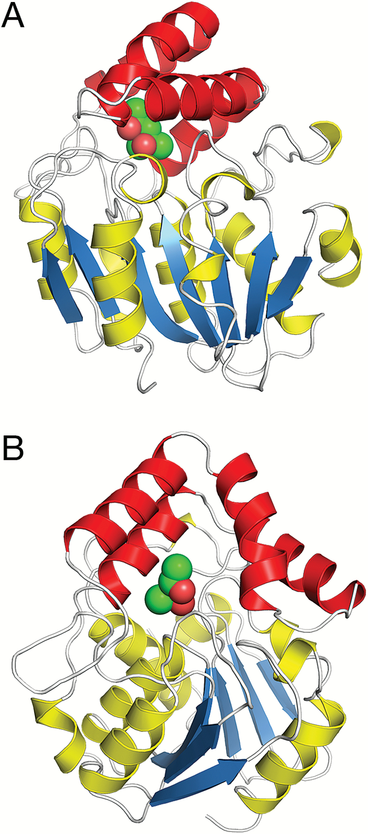
Fig. 1.
(A) Overall structure of OsD14. β-Strands of the core are coloured blue, associated helices yellow, and helices in the helical cap red. MPD is depicted as green spheres. (B) Binding of MPD in the ligand-binding cavity showing the opening of the cavity towards the solution. The two views in (A) and (B) are related by a 90° rotation with respect to the vertical axis.

