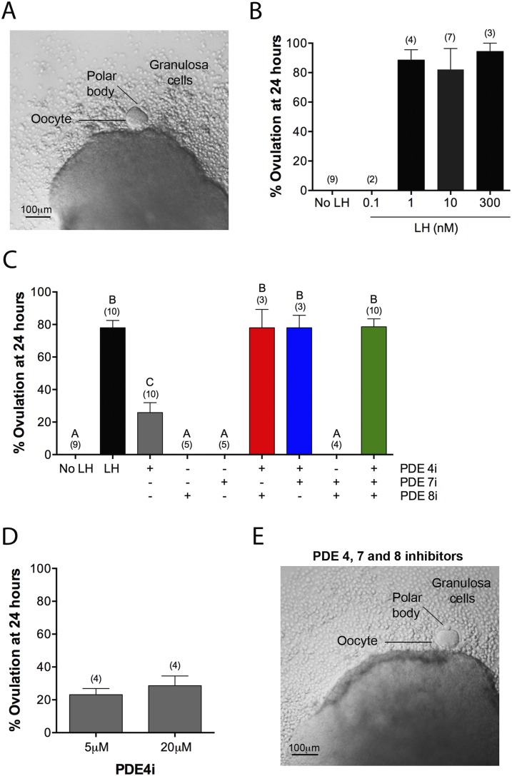
Figure 3.
PDE4, PDE7, and PDE8 act together to suppress premature ovulation. (A) A follicle at 24 hours after exposure to LH (300 nM). Ovulation has occurred, as indicated, by extrusion of the oocyte and some granulosa cells. (B) Percent ovulation at 24 hours after exposure to various concentrations of LH. (C) Percent ovulation at 24 hours after treatment of follicles with LH (300 nM), individual PDE inhibitors, pairs of inhibitors, or all three inhibitors together (5 μM rolipram, 1 μM PDE7i, 1 μM PDE8i). (D) Similar percent ovulation in response to 5 and 20 μM rolipram. (E) A follicle at 24 hours after treatment with a mixture of PDE4i, PDE7i, and PDE8i [concentrations as in (C)], showing ovulation, as seen with LH. Numbers in parentheses indicate the number of independent experiments. Different letters indicate significant differences (P < 0.05).

