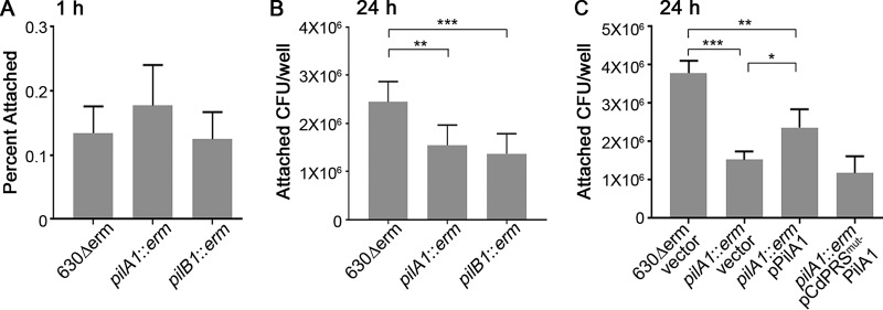
FIG 4.
Type IV pili promote adherence to MDCK cell monolayers at 24 h. (A and B) Attachment of 630Δerm and TFP-null mutants after 1 h (A) or 24 h (B) of incubation with MDCK cell monolayers. In panel A, data are expressed as the percentages of the original inoculum recovered following incubation and PBS washes, with 3 biological replicates for each strain. In panel B, data are expressed as the total CFU recovered per well after 24 h of incubation, with 6 biological replicates. Data were analyzed using a one-way ANOVA with Dunnett's test for multiple comparisons. (C) Complementation of the adherence defect of the pilA1 mutant after 24 h of incubation with MDCK cell monolayers. Symbols represent values from individual animals, and error bars indicate the standard deviations. Data were analyzed by one-way ANOVA using the Holm-Sidak method to correct for multiple comparisons. *, P < 0.05; **, P < 0.01; ***, P < 0.001.

