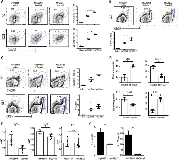
FIG 4.
T cell-intrinsic MyD88 deletion leads to more robust Tfh cell and GC responses upon B. burgdorferi infection but defective Th1 and Th17 responses. MyD88fl/fl and MyD88ΔT mice were infected with B. burgdorferi strain 297 (105 CFU/mouse) intradermally. (A) Proportion of CD4+ T cells in the spleen that express Tfh cell markers on day 14 postinfection and quantification of CD4+ Tfh cells from three independent mice. (B) CD19+ GL7+ CD95+ GC B cells in the spleens of infected mice on day 14 postinfection and quantification of GC B cells from three independent mice. (C) Proportion of Tfh cells and GCs as well as their quantification in the spleens of infected mice on day 21 postinfection. (D) Relative expression of the indicated genes in sorted CD4+ CD44hi CD62Llo cells on day 14 postinfection as quantified by qPCR. (E) Enzyme-linked immunosorbent assay quantification of B. burgdorferi-specific immunoglobulins in the serum of B. burgdorferi-infected MyD88fl/fl and MyD88ΔT mice on day 21 postinfection. (F) CD4+ CD44hi cells from the spleens of B. burgdorferi-infected mice were sorted on day 14 and cocultured with splenic DCs in the presence of 50 μg/ml B. burgdorferi extract. After 48 h of coculture, the supernatants were collected to measure IL-17A and IFN-γ. Data are representative of those from two to three independent experiments with three mice in each group. P values were determined by an unpaired t test. *, P > 0.05; **, P > 0.01; n.s., not significant.

