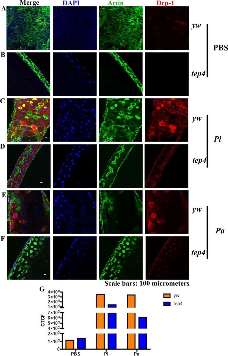
FIG 11.
Drosophila mutants for Tep4 display reduced DCP-1 expression in the midgut upon infection with Photorhabdus. (A to F) Guts from 7- to 10-day-old tep4 mutants and background control flies (yw) were stained with DCP-1 (red), DAPI (blue), and phalloidin (green). The dissected, stained tissues of flies injected with 1× PBS (A and B), P. luminescens (Pl) (C and D), and P. asymbiotica (Pa) (E and F) were viewed at a ×40 magnification by using confocal microscopy. (G) Corrected total cell fluorescence (CTCF) was measured to quantify the expression levels of DCP-1 in both background control flies (yw) and tep4 mutant flies by using ImageJ.

