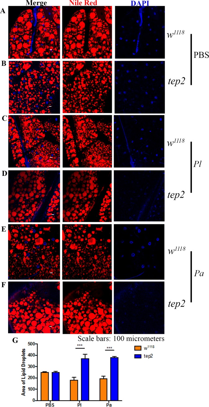
FIG 5.
Drosophila mutants for Tep2 display large lipid droplets after Photorhabdus infection. (A to F) Fat body tissues were stained with Nile Red-O as well as DAPI (4′,6-diamidino-2-phenylindole) and observed under a confocal microscope (Olympus) at a ×20 magnification. Lipid droplets (red) and nuclei (blue) are shown for flies of the background control strain (w1118) (A, C, and E) and the tep2 mutant strain (B, D, and F) 18 h after infection with Photorhabdus luminescens (Pl) or P. asymbiotica (Pa) or injection with 1× PBS (negative control). (G) Areas of lipid droplets in fat body cells of the background control strain (w1118) as well as tep2 mutants were quantified by using ImageJ. The means from at least three independent fat body samples are shown, and error bars represent standard deviations. Significant differences are shown with asterisks (***, P < 0.001).

