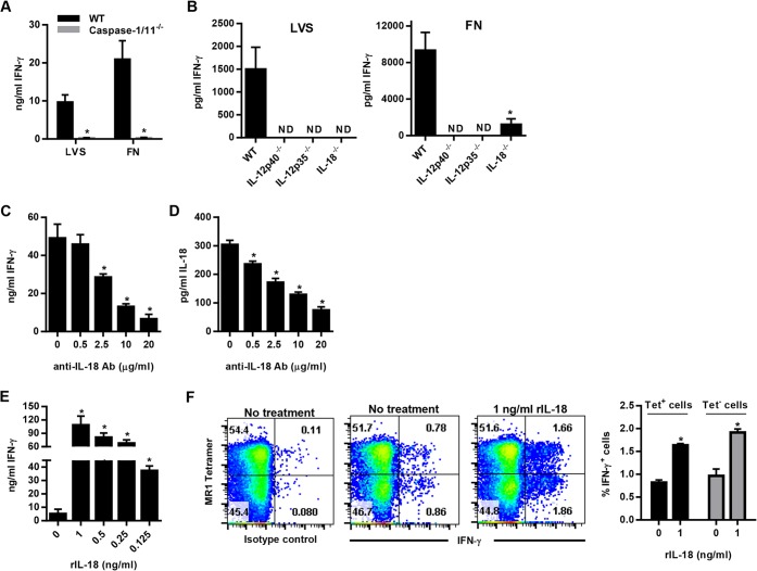
FIG 4.
IL-18 modulates the level of MR1-5-OP-RU tetramer+ and tetramer− Vα19iTg T cell IFN-γ responses to F. tularensis LVS and F. novicida. (A) WT and caspase-1/11−/− macrophages were infected with either LVS or F. novicida (FN) at an MOI of 1:1 and cocultured with Vα19iTg T cells. IFN-γ was measured in the supernatants after 20 h of culture. *, P < 0.01 compared to WT macrophages infected with the same organism. (B) WT, IL-12p35−/−, IL-12p40−/−, and IL-18−/− macrophages were infected with either LVS or F. novicida (FN) at an MOI of 1:1 and cocultured with Vα19iTg T cells. IFN-γ was measured in the supernatants after 20 h of culture. *, P < 0.01 compared to WT macrophages infected with the same organism. (C) WT macrophages were infected with F. novicida at an MOI of 1:1 and cocultured with Vα19iTg T cells. Neutralizing anti-IL-18 Ab was added at the indicated concentrations at the start of coculture, and IFN-γ was measured in the supernatants after 20 h of culture. *, P < 0.01 compared to cultures containing no anti-IL-18 Ab. (D) Levels of IL-18 measured in the supernatants of cultures depicted in panel C. *, P < 0.01 compared to cultures containing no anti-IL-18 Ab. (E) WT macrophages were infected with F. tularensis LVS at an MOI of 1:1 and cocultured with Vα19iTg T cells. Recombinant IL-18 was added at the indicated concentrations at the start of coculture, and IFN-γ was measured in the supernatants after 20 h of culture. *, P < 0.01 compared to cultures containing no recombinant IL-18. (F) IFN-γ intracellular cytokine staining of Vα19iTg T cells cultured with F. tularensis LVS-infected WT macrophages for 20 h in the presence of 1 ng/ml recombinant IL-18, or no additional IL-18 (“no treatment”), as indicated. The percentages of intracellular IFN-γ+ cells for the 5-OP-RU MR1 tetramer+ and tetramer− populations are depicted graphically. *, P < 0.01 compared to cultures containing no additional recombinant IL-18. Brefeldin A was added during the final 4 h of culture. All flow cytometry dot plots depict TCRβ+ lymphocytes with B220+ and F4/80+ cells excluded by electronic gating. No treatment, culture medium without recombinant cytokines; isotype control, intracellular cytokine staining using a nonspecific control Ab matched to the isotype of the IFN-γ Ab. Data represent values ± SEM from three replicates and are representative of three independent experiments.

