FIG 5.
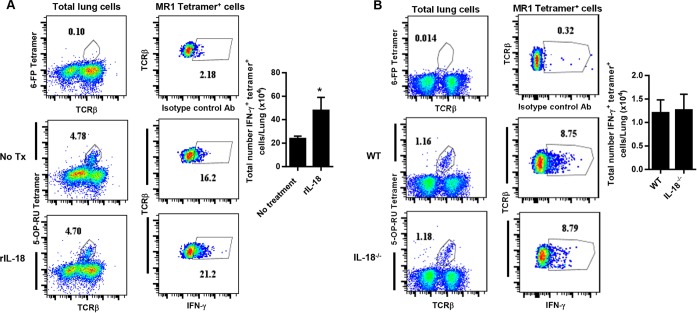
MR1-5-OP-RU tetramer+ T cell IFN-γ responses during in vivo F. tularensis LVS pulmonary infection. (A) Wild-type C57BL/6 mice were infected with a sublethal intranasal dose of 2 × 10² CFU F. tularensis LVS and administered supplemental recombinant IL-18 (rIL18) delivered intranasally on days 7 and 10. IFN-γ intracellular cytokine staining of 5-OP-RU MR1 tetramer+ TCRβ+ cells in the lungs of mice given supplemental rIL-18 or PBS (no treatment) was assessed on day 11 after infection. (B) Wild-type C57BL/6 mice and IL-18−/− mice were infected with a sublethal intranasal dose of 2 × 10² CFU F. tularensis LVS. IFN-γ intracellular cytokine staining of 5-OP-RU MR1 tetramer+ TCRβ+ cells was assessed in the lungs on day 8 after infection. Control tetramer (6-FP) staining is shown for comparison. The total numbers of intracellular IFN-γ+ 5-OP-RU MR1 tetramer+ cells are depicted graphically. *, P < 0.01 compared to control mice. Data represent values ± SEM from 3 to 5 individual mice and are representative of 3 or 4 independent experiments.

