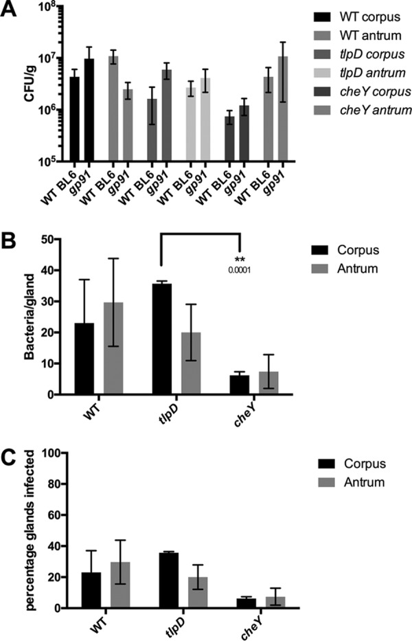
FIG 4.

Loss of immune superoxide rescues tlpD mutant gland defects. Colonization of Cybb−/− mice by WT, tlpD mutant, or cheY mutant GFP+ H. pylori SS1 strains at 2 weeks postinfection is shown. Mice were orally infected, and stomachs were collected and analyzed for tissue and gland colonization. (A) Numbers of CFU/g at 2 weeks postinfection for corpus and antrum regions using WT (n = 6), tlpD mutant (n = 14), and cheY mutant (n = 6) GFP+ H. pylori SS1 strains. Data for WT mice are the same as those in Fig. 1 and are displayed here for comparison. WT BL6 refers to wild type and gp91 refers to Cybb−/− mice. (B) Gland loads in the isolated corpus and antral glands, representing the average number of bacteria counted per gland, excluding uninfected glands. Numbers of infected glands are as follows: WT-infected corpus, 69 glands from three mice; WT-infected antrum, 89 glands from three mice; tlpD mutant-infected corpus, 107 glands from three mice; tlpD mutant-infected antrum, 60 glands from three mice; cheY mutant-infected corpus, 31 glands from three mice; cheY mutant-infected antrum, 37 glands from three mice. (C) Gland occupancy in the isolated corpus and antral glands, representing the percentage of glands infected with the indicated H. pylori strain. Error bars represent SEM for all panels. Numbers of mice infected are the same as described for gland loads. Statistical differences are indicated by * (P < 0.05) and ** (P < 0.01) as analyzed by Student t test.
