FIG 1.
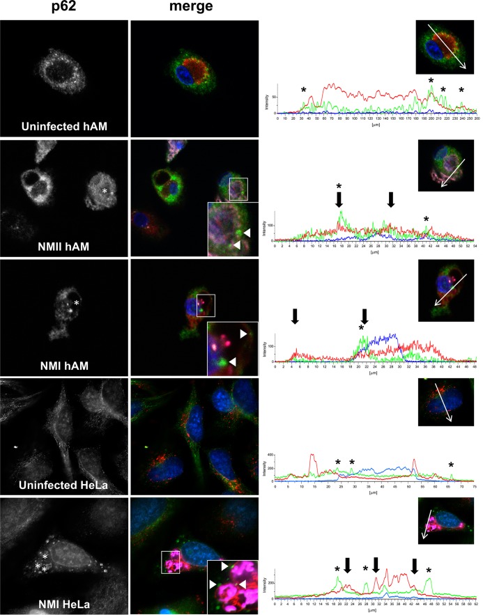
p62 localizes near the PV during primary hAM infection with C. burnetii NMI and NMII. (Left and middle) hAMs or HeLa cells were infected for 72 h with virulent (NMI) or avirulent (NMII) C. burnetii or left uninfected and then processed for immunofluorescence confocal microscopy to detect p62 (green), CD63 (red), and C. burnetii (violet). DAPI-stained DNA is blue. The data shown are representative of observations from three individual donors and experiments. (Insets) Magnified regions of p62 localization around the PV. Arrowheads, p62 puncta that colocalize with CD63; asterisks in p62 panels, PV. (Right) Intensity profiles show the limiting membrane of the PV (arrows) and the peaks of p62 intensity (asterisks) near the PV membrane. Native p62 localizes near the CD63-positive PV in primary hAMs and HeLa cells infected with virulent C. burnetii.

