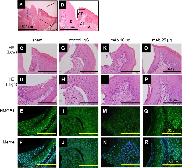
FIG 2.
Immunofluorescence localization of HMGB1 in periodontitis mice. (A) Histological image from a healthy mouse (sham) at day 7 (low magnification). Bar, 500 μm. (B) Enlargement of the section of the image indicated in panel A. D, dentin; GE, gingival epithelium; CT, connective tissue; A, alveolar bone. Bar, 200 μm. We performed each staining at least three times. Images of the gingival junctional epithelium for HE and immunofluorescence of HMGB1 (green) and DAPI (blue) in sham and periodontitis mice are shown as follows: sham, panels C to F; control IgG administration group, panels G to J; anti-HMGB1 antibody administration group, panels K to N (10 μg/mice) and O to R (25 μg/mice). Merged images (F, J, N, and R) indicating colocalization are also shown. Bars, 100 μm (low magnification) (C to O) and 50 μm (high magnification) (D to R).

