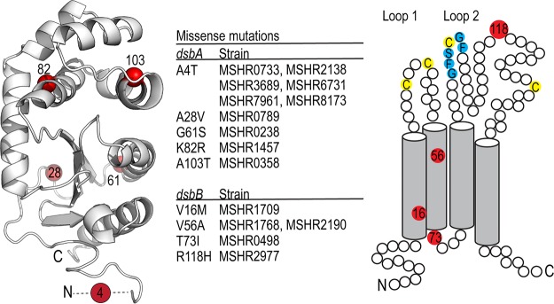
FIG 1.
DsbA and DsbB are highly conserved among a diverse collection of B. pseudomallei isolates. Analysis of the genetic variation of DsbA and DsbB among 431 clinical isolates of B. pseudomallei sourced from the Darwin Prospective Melioidosis Study found both genes to be highly conserved. The vast majority of isolates possess a sequence identical to that of strain K96243, with only a few mutations being observed in a limited number of strains (center). The observed five missense mutations (Ala4Thr, Ala28Val, Gly61Ser, Lys82Arg, and Ala103Thr) observed for BpsDsbA are mapped onto the crystal structure of BpsDsbA (PDB accession number 4K2D, 1.9-Å resolution) (left). Mutation positions are colored red, and the relevant C-α atom is shown as a sphere. The 6 most N-terminal residues could not be resolved in the electron density of this crystal structure and are represented by a dashed line. For BpsDsbB, we observed four missense mutations (Val16Met, Val56Ala, Thr73Ile, and Arg118His), again in only a few of the 431 isolates (center). The positions of the mutations (red circles) are depicted in a schematic of BpsDsbB. Transmembrane helix boundaries were predicted using the TMHMM server (76, 77). Key cysteine residues in each periplasmic loop and the predicted BpsDsbA interaction sequence Gly-Phe-Ser-Cys-Gly-Phe (Fig. 5) are marked in blue.

