
Fig. 10.
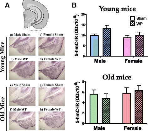
Detection of epigenetic DNA modifications with 5-hmC immunohistochemistry. A Paraformaldehyde-fixed brains from young (a–d) and old (e–h) brain tissues were coronally frozen-sectioned at 40 μm and were stained with an anti-5-hmC antibody. Immunoreactivity in the area including the temporal lobe and amygdala (dotted rectangle in the top diagram) was qualitatively evaluated in sham (a, c, e, g) and WP-sensitized (b, d, f, h) mice. Representative photomicrographs were taken using a ×4 objective (scale bar = 0.5 mm). B Immunoreactivity to 5-hmC (5-hmC-IR) within the young and old mouse brains was quantified by densitometric analyses of the digital photomicrographs taken with a ×4 objective. The values indicate group average optical density ± standard error (n = 5–6). Top graph, young mice; bottom graph, old mice