
Fig. 4.
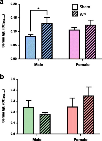
Assessment of WP-specific IgE levels in the sera from sham and WP-sensitized mice using ELISA. Relative levels of WP-specific IgE were determined in the sera from sham or WP-sensitized young (a) and old (b) male and female mice. Each serum sample was diluted 1:1 with assay buffer prior to the assay. The amounts of WP-specific IgE were determined by the colorimetric substrate reaction and the average optical density (OD) at 450 nm for the experimental groups were compared (average OD ± standard error). The open bars and hashed bars indicate sham and WP-sensitized groups, respectively. Young male, n = 7–8; young female, n = 8; old male, n = 6; old female, n = 7, *p < 0.05