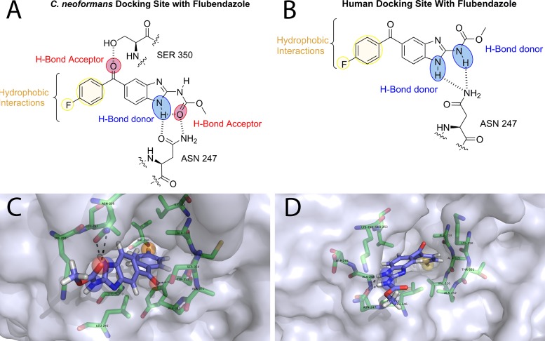
FIG 1.
(A and B) Homology model of flubendazole docked with both C. neoformans (A) and human (B) β-tubulin. Red sphere, hydrogen bond donors; blue sphere, hydrogen bond acceptors; yellow sphere, hydrophobic interactions. (C and D) The docking pose is visualized with PyMOL. Protein is shown as a surface representation colored 40% transparent light blue. Flubendazole is represented as sticks composed of carbon (light blue), hydrogen (white), nitrogen (dark blue), oxygen (red), and fluorine (cyan). Binding site residues selected around 4 Å are represented as sticks with carbon (green), nitrogen (blue), oxygen (red), and sulfur (yellow).

