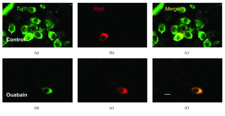
Figure 3.
Images of spiral ganglion neurons in control and ouabain-treated ears. (a–c) Representative image of Tuj1 and Prph immunostaining in spiral ganglion cells of the control group. Prph, a marker of type II spiral ganglion cells, was detected with Alexa Fluor 594 (red). Tuj1, a marker of type I and type II neurons, was detected with Alexa Fluor 488 (green). (d–f) Representative image of Tuj1 and Prph immunostaining in spiral ganglion cells of the ouabain-treated group. Nearly, all type I spiral ganglion cells were lost, whereas Prph-positive type II neurons survived after ouabain exposure. Scale bar = 20 μm.

