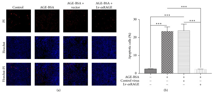
Figure 2.
Effects of esRAGE on AGEs associated HUVEC apoptosis. Endothelial cells were cultured with complete medium containing 200 μg/mL AGE-BSA for 24 h, submitted to the Hoechst-PI double-staining method, and photographed under a fluorescence microscope using the ZEN software. Total and apoptotic cells were counted using the Image-Pro-Plus 6.0 software, based on which apoptotic rates were calculated. (a) Cell staining in the 4 HUVEC groups, imaged by fluorescence microscope after Hoechst-PI staining. (b) Apoptotic rates in the 4 HUVEC groups. ∗∗∗P < 0.01.

