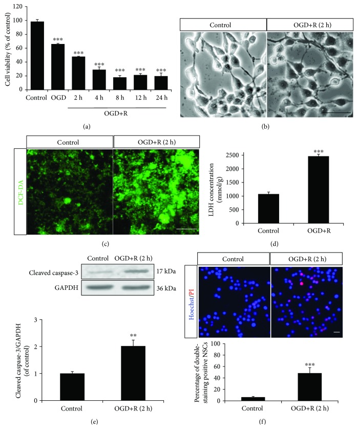
Figure 1.
OGD/reoxygenation-induced NSC apoptosis. The NSCs cultured in the monolayer were subjected to OGD for 2 hours and reoxygenation for 2, 4, 8, 12, and 24 hours of induction. (a) Cell viability decreased relating to the duration of OGD/reoxygenation treatment through MTT assay. (b) The NSCs displayed an unhealthy morphology following OGD/reoxygenation induction. (c) The NSCs produced extra ROS with OGD/reoxygenation induction, according to DCF-DA fluorescence probe labelling. (d) The LDH of NSCs increased with OGD/reoxygenation induction, according to cellular LDH detection. (e) The protein expression of cleaved caspase-3 in NSCs was upregulated with OGD/reoxygenation induction, according to Western blotting. (f) OGD/reoxygenation treatment increased apoptosis of NSCs (Hoechst 33342/PI, double staining positive cells). ∗∗P < 0.01 and ∗∗∗P < 0.001 were considered to be significantly different between control and OGD or control and OGD+R groups. n = 3. Scale bar: 20 μm.

