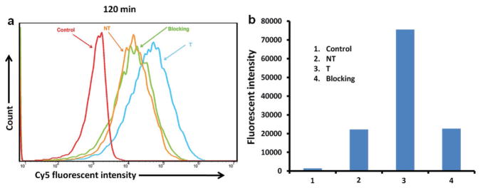
Figure 4.
Cellular uptake of the unimolecular nanoparticles in ovarian cancer cells. OVCAR-3 cells were treated with medium (Control), targeted (T), or nontargeted (NT) unimolecular nanoparticles, or a combination of targeted nanoparticles and free cRGD (i.e., the blocking assay) for 2 h. A) The fluorescence intensity of Cy5 as represented by flow cytograms. B) The relative fluorescence intensity analyzed by FlowJo 10.0.5 analysis software.

