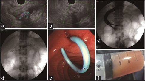
Figure 3.

The second emergency endotherapy treatment images. (a) The endoscopic ultrasonography showed the obviously dilated minor pancreatic duct with a maximum diameter of 25 mm, obstructed by multiple stones and the largest one measured 12 mm × 6.8 mm. (b) The endoscopic ultrasonography-guided puncture of minor pancreatic duct was performed with a 19-gauge needle through the duodenal bulb. (c) The puncture passage was dilated by the 7F Soehendra stent retriever. (d) An 8 cm 7F plastic stent was implanted into minor pancreatic duct. (e) The stent located well and the pancreatic juice flowed out fluently. (f) 40 ml cloudy pancreas juice was extracted
