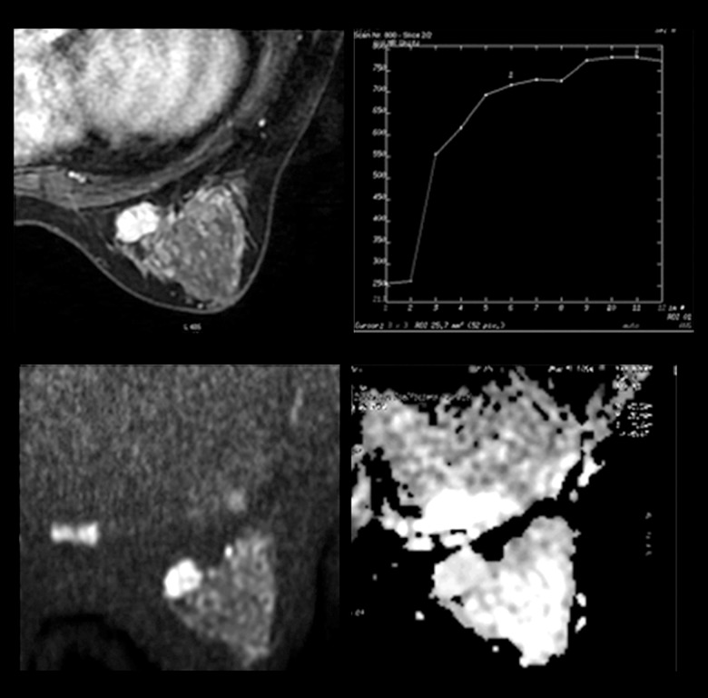
Figure 2.
MRI of a 40-year-old patient showing a lesion in the left breast. Dynamic contrast-enhanced images revealed an oval mass enhancement with in-heterogeneous internal enhancement, and a continuous increasing curve type. Initial enhancement was 120% and the ADC value was 1.77×10⁻³ mm2/s. The lesion was rated as BI-RADS Category 4A. The pathology was fibroadenomas.

