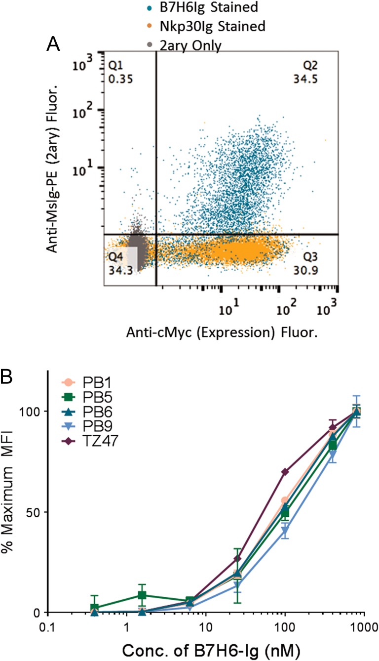
Fig. 2.
Characterization of Yeast-Displayed PB Clones. (A) Population g2.4 was stained with B7H6-Ig, Nkp30-Ig and secondary only using an anti-MsIg-PE secondary for fluorescence read-out (y-axis). Yeast were also stained for the cMyc expression tag (x-axis). (B) A range of B7H6-Ig antigen concentrations were used to stain yeast-displayed PB clones and TZ47. MFI values were normalized to represent the % of the maximum signal observed at the highest concentration tested. Error bars indicate the standard deviation of three technical replicates and relative trends are representative of three experimental replicates.

