Fig. 6.
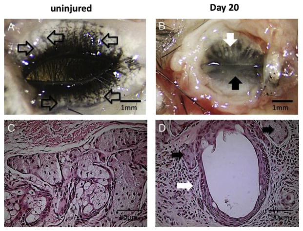
A. Eyelids of a C57BL/6 mouse. Eyelids were excised and observed from the behind. Acini were observed (open arrows). B. Meibomian glands of a C57BL/6 mouse 20 day s after ocular surface alkali burn. Loss of the acini and dilated ducts were observed in the upper eyelid (white arrow). In the lower eyelid meibomian gland was not observed (black arrow). C. Histology of meibomian glands of a C57BL/6 mouse. D. Histopathology of meibomian glands in the upper eyelid of a C57BL/6 mouse 20 days after ocular surface alkali burn. Largely dilated duct (open arrow) of the meibomian glands is observed, acinar cells (black arrows) are also seen among hypercellular mesenchyme (reproduced from Mizoguchi, 2015).

