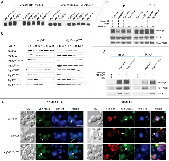
Figure 7.

Mutations in Atg30 affect pexophagy, Atg30 phosphorylation and Atg11 recruitment. (A) Phosphorylation status of Atg30 WT and different Atg30 mutants expressed in atg30Δ or atg30Δ atg37Δ cells (sKZR009-20 and sKZR030-31; Table S3) was analyzed by western blot after 15 h of methanol induction. (B) Pexophagy assay for Atg30 mutants. Methanol-grown cells expressing Atg30 WT or different Atg30 mutants expressed in atg30Δ and atg30Δ atg37Δ cells (sKZR009-20 and sKZR030-31; Table S3) were adapted to SD-N medium. Cell lysates were prepared as described in Materials and Methods and resolved by SDS-PAGE. Western blotting was performed with antibodies against P. pastoris Aox1. (C) Atg30Y236A, Atg30L241A and Atg30Δ236-244 mutants co-immunoprecipitate with Atg11. Methanol-grown ypt7Δ cells expressing FLAG-Atg11 and Atg30WT, Atg30Y236A, Atg30L241A or Atg30Δ236-244 mutants (sKZR026-29 - see Table S3), were adapted to SD-N medium for 0.5 h to induce pexophagy before the co-IP:HA experiment was conducted. Immunoprecipitated proteins were visualized with anti-HA, anti-FLAG and anti-P. pastoris Pex3 antibodies. (D) The Atg30W210,212A mutant has severely reduced ability to co-immunoprecipitate with Atg37, but not with Pex3. Methanol-grown ypt7Δ atg30Δ cells expressing GFP-Atg37 and cells expressing GFP-Atg37 and Atg30 WT or the Atg30W210,212A mutant (sTN566, sKZR021and sKZR022; Table S3), were adapted to SD-N medium for 0.5 h to induce pexophagy before the co-IP:HA experiment was conducted. Immunoprecipitated proteins were visualized with anti-HA, anti-GFP and anti-P. pastoris Pex3 antibodies. (E) Interaction between Atg37 and Atg30 is required for localization of Atg11 in close proximity to peroxisomes. Methanol-grown Atg30 WT, atg30Δ and Atg30W210,212A cells expressing BFP-SKL and GFP-Atg11 (sTN640, sKZR023and sKZR024; Table S3) were adapted to SD-N for 15 min or 1 h and stained with FM 4-64 for vacuole visualization. White arrowheads indicate GFP-Atg11 localization. Scale bars: 5 µm.
