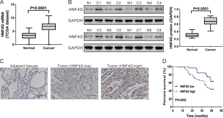
Figure 1. Expression of HNF4G in lung cancer tissues and normal lung tissues.
(A) HNF4G mRNA expression analysis based on TCGA dataset, which included 58 normal lung tissues and 488 lung cancer tissues. (B) Western blotting analysis of HNF4G and FOXO3 protein in tissue samples. Representative blot (left panel) and quantification of three independent experiments (right panel) were shown. T1-T8, tumor tissue, C1-C8, adjacent normal lung tissue. (C) Expression of HNF4G was determined by immunohistochemical staining in lung cancer and adjacent normal tissues (n = 85). Magnification: 400´. Scale bars: 50 μm. (D) Kaplan–Meier survival analyses of 85 patients with lung cancer. Survival analysis showed that HNF4G-low expression tumors (n = 32) had a favorable prognosis compared to HNF4G-high expression tumors (n = 53) (P < 0.01).

