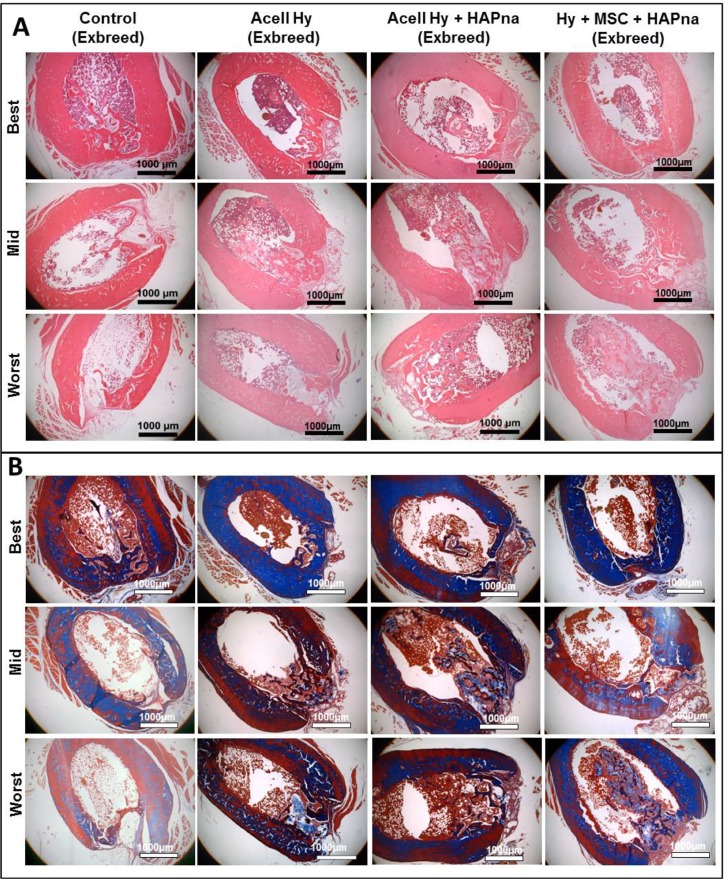
Figure 4.
Histological assessment of the defect site after 4 weeks repair time following a non-critical sized defect in the midshaft of the femur in exbreeder female (>6 months old) rats, stained with H&E (A) or Masson's trichrome (B). Representative images from 6 replicates for each experimental group to demonstrate the best, mid and worst bone repair observed from independent pathological assessment. Scale bar: 1000 μm.

