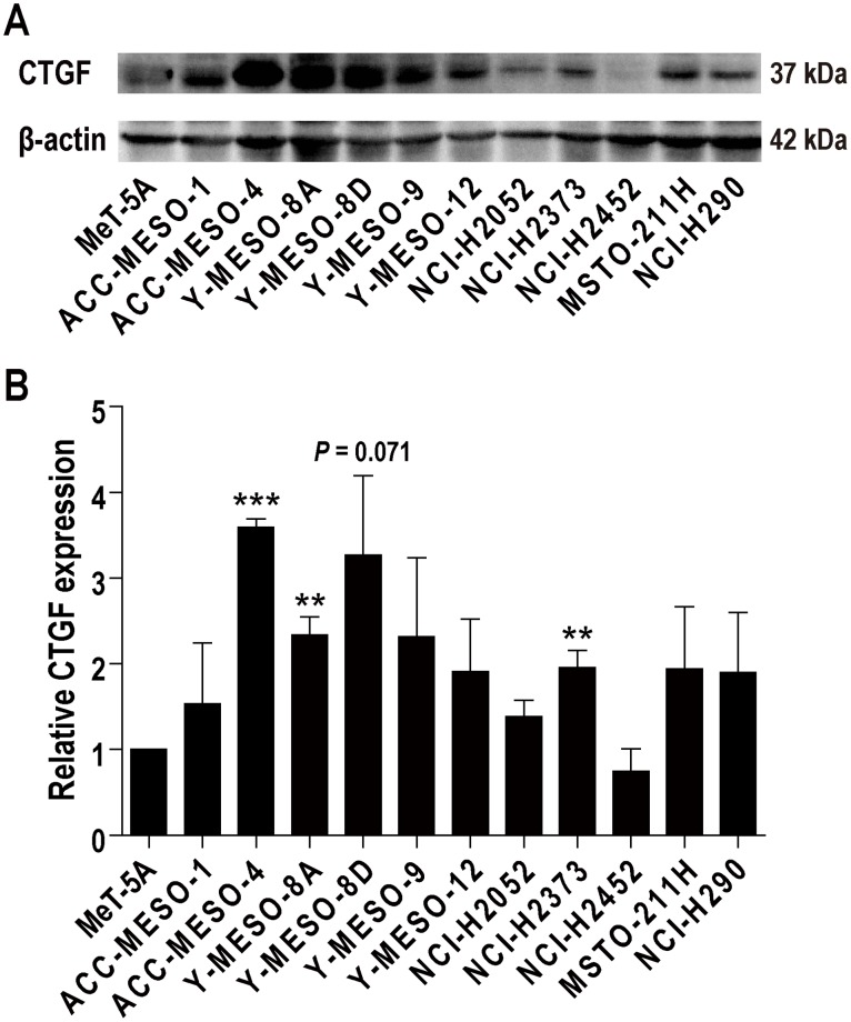
Figure 1. CTGF expression in human mesothelioma cell lines.
(A) Western blot analysis. Antibody 14939 (Santa Cruz Biotechnology; 1:200) was used to detect CTGF at 36-38 kDa. All the cell lines examined expressed CTGF, but several cell lines expressed low levels of CTGF, irrespective of histological subtypes. Three cell lines (ACC-MESO-4, Y-MESO-8D and NCI-H290) were chosen for the following experiments. ACC-MESO-4 and NCI-H290 are epithelioid subtype, and Y-MESO-8D is sarcomatoid subtype. (B) Semiquantitative analysis of western blot analysis. Relative CTGF expression in comparison to MeT-5A was calculated with ImageJ. N = 3; means ± SEM, **P < 0.01, ***P < 0.001.

