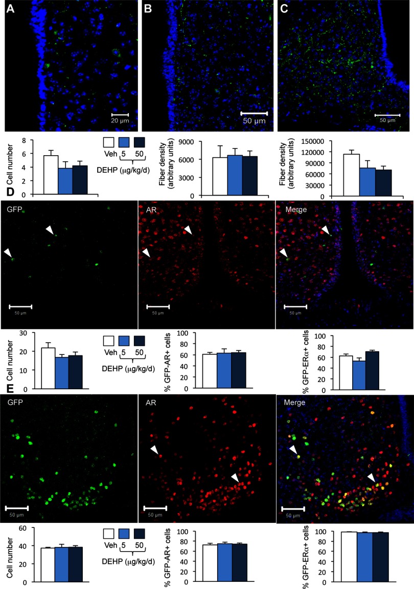
Figure 4.
Effects of di(2-ethylhexyl) phthalate (DEHP) on kisspeptin, green fluorescent protein (GFP)/androgen receptor (AR) and immunoreactivity in wild type and Kiss1-creGFP male mice. Mice were treated with DEHP (0.5, 5, or ) or the vehicle (Veh). Analyses were performed in the rostral periventricular area of the third ventricle (RP3V) and arcuate nucleus. (A–C) Representative immunolabelling (upper panels) and quantification (lower panels) of the number of kisspeptin-immunoreactive neurons in the RP3V (A) and fiber density in the RP3V (B) and arcuate nucleus (C) in wild type mice. Data are expressed as the means of six males per treatment group. (D) Upper panels: Representative immunolabelling of GFP- (left), AR-immunoreactivity (middle), and the merge (right) in the RP3V of Kiss1-creGFP males. Lower panels: quantification of the number of GFP-cells (left), GFP/AR- (middle), and -coexpressing cells (right). Data are expressed as the means of five to six males per treatment group. (E) Upper panels: Representative immunolabelling of GFP- (left), AR-immunoreactivity (middle), and the merge (right) in the arcuate nucleus of Kiss1-creGFP males. Lower panels: quantification of the number of GFP-cells (left), GFP/AR-(middle), and -coexpressing cells (right). Data are expressed as the means of five to six males per treatment group.

